A spine T-score compares your lumbar bone density to that of a healthy young adult, and the number tells you which side of a clinical line you fall on: -1.0 and above is normal, between -1.0 and -2.5 is low bone density, and -2.5 or lower is osteoporosis. Clinicians rarely stop at the number, though. They pair it with the FRAX tool and your fracture history before deciding on treatment.
TL;DR:
- Spine T-scores above -2.5 can be falsely elevated due to osteophytes, aortic calcification, or vertebral fractures, which can mislead diagnosis.
- The spine is more sensitive to early bone loss because of its higher trabecular bone content but is also more prone to artifacts affecting accuracy.
- A T-score of -2.5 or lower indicates osteoporosis, but fracture risk assessment with FRAX and clinical history remains essential for treatment decisions.
- Lifestyle factors like calcium intake, exercise, smoking, and fall prevention can influence bone density, but genetics and age are fixed risks.
- Pharmacologic treatments reliably improve spine density over years, while loading therapies are adjunct options requiring clinician supervision.
Table of Contents
- What a spine T-score measures and how DXA produces it
- How to interpret spine T-score numbers and how FRAX informs treatment decisions
- Why spine DXA results can be misleading
- When spine T-score is useful and when hip or CT may be better
- What affects your spine T-score and modifiable actions that matter
- Can your spine T-score improve? Evidence and realistic expectations
- Next steps after an abnormal spine T-score
- Integrating T-score data into an individualized care plan
- A supervised option worth discussing with your provider
- Sources
- FAQ
What a spine T-score measures and how DXA produces it
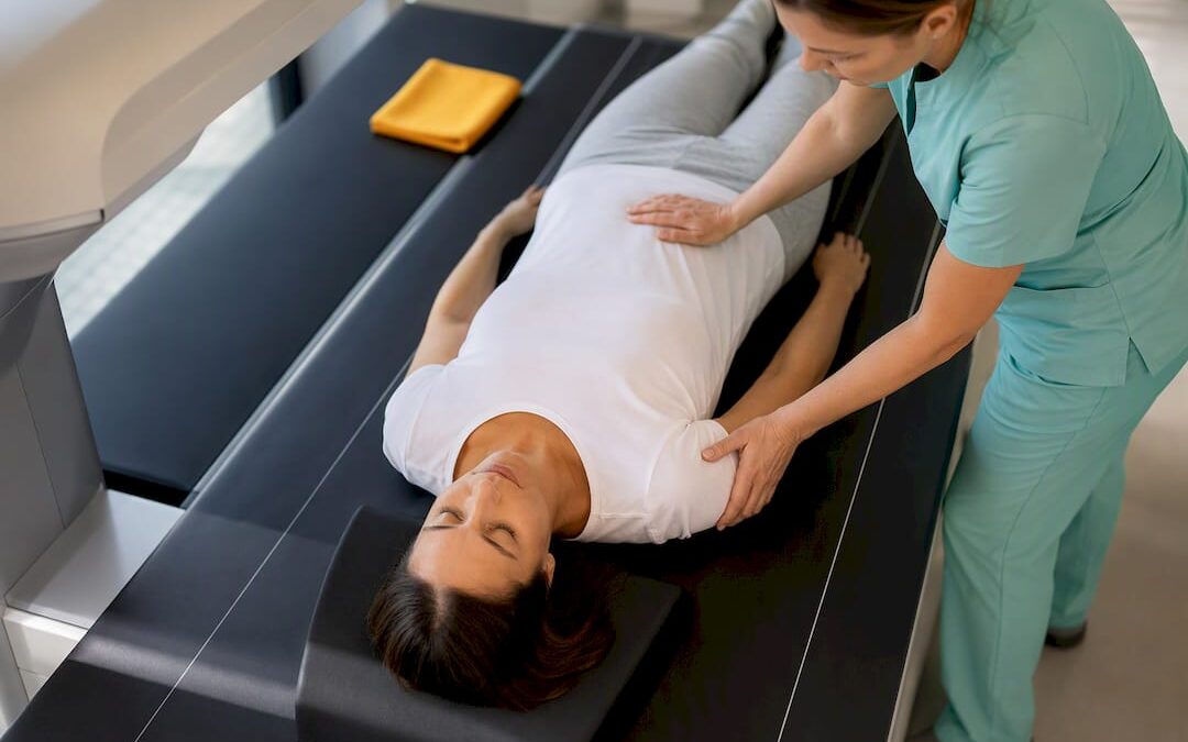
A dual-energy X-ray absorptiometry scan, usually shortened to DXA, sends two low-dose X-ray beams through your bone and soft tissue to calculate mineral density. The scanner reports two comparisons for the same measurement, and the difference between them matters.
Your T-score stacks your bone density against a healthy young adult of the same sex at peak bone mass, which is why it drives the diagnostic categories doctors use. A Z-score instead compares you to others your own age, sex, and often ethnicity, and clinicians lean on it for children, premenopausal women, and younger men where age-matched context tells a more useful story than a comparison to peak bone mass ever could.
DXA typically scans two regions:
- The lumbar spine, usually averaging vertebrae L1 through L4
- The hip, including the femoral neck and total hip
These sites get chosen because they carry a mix of trabecular and cortical bone and because fractures there carry the heaviest health consequences, according to MedlinePlus.
How to interpret spine T-score numbers and how FRAX informs treatment decisions
The numeric categories are consistent across major health authorities. A T-score of -1.0 or higher is normal bone density. A score between -1.0 and -2.5 signals low bone density, often called osteopenia. A score of -2.5 or lower meets the threshold for osteoporosis.
- Normal: T-score of -1.0 or above
- Low bone density (osteopenia): T-score between -1.0 and -2.5
- Osteoporosis: T-score of -2.5 or below
A T-score alone rarely settles the treatment question, though. A fragility fracture, meaning a break from a fall at standing height or less, often triggers treatment regardless of what the scan shows, because a prior fracture predicts future fracture risk on its own.
FRAX combines your T-score with clinical risk factors to estimate your 10-year fracture probability. Practices commonly treat when the FRAX-calculated hip fracture risk is considered elevated or when the major osteoporotic fracture risk is deemed significant by clinical judgment. A clinician familiar with your history calculates and interprets this score, since age, prior fracture, smoking, glucocorticoid use, and family history all feed into it.
Why spine DXA results can be misleading
The spine is prone to false readings in ways the hip usually is not. Several common conditions inflate the measured density without reflecting a real gain in bone strength, and the International Society for Clinical Densitometry has built specific rules to catch them.
- Osteophytes, the bony spurs that form with osteoarthritis, add extra mineral to the scan area and push the T-score artificially higher.
- Aortic calcification sits directly over the lumbar spine on many scans and can inflate the reading the same way.
- Vertebral compression fractures distort the shape of the vertebra and can skew results in either direction.
ISCD guidance calls for excluding any vertebra with a visible structural abnormality or one whose T-score differs by more than 1.0 standard deviation from its neighbors. If exclusions leave only one usable vertebra, the spine T-score should not be used for diagnosis at all.
Pro Tip: Ask your clinician directly whether any vertebrae were excluded from your spine reading, and whether a hip DXA or vertebral imaging was done to confirm the result.

When spine T-score is useful and when hip or CT may be better
The spine has an advantage early in bone loss: it contains a higher proportion of trabecular bone, the more metabolically active tissue that tends to lose density before cortical bone does. That sensitivity makes it useful for catching change sooner, but it is the same tissue structure that makes it vulnerable to the artifacts described above.
- Spine T-score often flags early bone loss before hip T-score does.
- Hip T-score tends to better predict fracture risk at non-spine sites and is favored when spine imaging shows degenerative change.
- CT-based assessment using Hounsfield units can offer a more localized look at a specific vertebra, which researchers have explored as a preoperative option when standard DXA readings look unreliable, according to research comparing CT and DXA.
When your spine and hip scores disagree, most clinicians weigh the hip more heavily for overall fracture risk.
What affects your spine T-score and modifiable actions that matter
Some drivers of bone density are fixed. Age, sex, genetics, and a prior fracture all shape your starting point and cannot be changed. Others respond to what you do.
- Calcium and vitamin D intake support the mineralization process bones depend on.
- Resistance and impact exercise stimulate bone remodeling in ways that low-impact activity does not.
- Smoking and heavy alcohol use both interfere with bone formation.
- Fall prevention, including vision checks, home safety, and balance training, reduces fracture risk independent of density itself.
Osteoporosis often causes no symptoms until a fracture occurs, which is a central reason NIAMS frames screening and early action as the practical defense.
Medication falls into two broad categories. Antiresorptive drugs, such as bisphosphonates, slow the rate of bone breakdown. Anabolic agents work differently, stimulating new bone formation directly. Both categories can raise spine BMD on repeat scans, but the timeline is measured in years, not weeks, and the size of the change depends on the drug class and your baseline density.
Can your spine T-score improve? Evidence and realistic expectations
Pharmacologic treatment is the intervention most reliably tied to measurable spine BMD gains, though the size of that gain varies by drug class and by how low your starting density was. Lifestyle changes and structured loading programs can add modest gains over time, but they work on a slower curve than medication does.
- Repeat DXA scans typically happen every one to two years to track meaningful change.
- Small shifts between scans can fall within the machine’s own measurement error rather than reflecting a real biological change.
- Clinicians use a calculated least significant change threshold to judge whether a shift in your number is real or just noise, a concept detailed in ISCD’s measurement guidance.
That precision issue matters for how you read your own results. A T-score that moves from -2.3 to -2.1 over a year might represent genuine improvement, or it might sit inside the scanner’s own margin of error depending on your facility’s specific precision data. Ask your provider what threshold they use before reading too much into a small change.
Next steps after an abnormal spine T-score
- Ask whether any vertebrae were excluded from your spine reading and why.
- Ask for your FRAX score and how it factored into the treatment recommendation.
- Ask about screening for secondary causes, such as thyroid or parathyroid conditions, that can drive bone loss.
- Discuss whether a hip DXA, a vertebral fracture assessment, or blood work makes sense as a follow-up.
Recent severe back pain, measurable height loss, or a known vertebral fracture are red flags that warrant prompt evaluation rather than waiting for your next scheduled scan.
Integrating T-score data into an individualized care plan
A spine T-score is one data point, not a verdict. The number means something different next to a clean scan than it does next to visible degenerative change, and it means something different in a person with a prior fracture than in someone without one.
The most useful conversations combine the T-score, the FRAX estimate, and your own fracture and medical history, then let you and your clinician weigh the options together.
— Aaron
A supervised option worth discussing with your provider
If you are looking for a non-drug addition to your care plan, osteogenic loading is a supervised, in-clinic session done once a week, designed to place brief, targeted mechanical stress on bone to stimulate new growth. It is meant to sit alongside your medical evaluation, not replace medications your clinician has indicated for you.
- Sessions run once weekly and are supervised.
- The center also offers red light therapy, PEMF mats, and other supportive therapies as adjuncts.
- The approach is non-pharmaceutical and is presented as a complement to standard care.
| Offering | Frequency | Role in care |
|---|---|---|
| Osteogenic loading | Once weekly | Adjunct to medical treatment |
| Red light therapy | As scheduled | Supportive therapy |
| PEMF mats | As scheduled | Supportive therapy |
Bring your T-score and FRAX results to a conversation with your provider before adding any new therapy, and check current locations to see what is available near you.
Sources
For readers who want to go deeper or bring material to a clinician, these sources back the figures and rules cited above:
- Bone Mineral Density Tests: What the Numbers Mean | NIAMS
- Bone Density Scan: MedlinePlus Medical Test
- International Society for Clinical Densitometry (ISCD)
- CT vs DXA comparisons and vertebral subregion BMD research (PMC)
This article is general information, not a substitute for advice from a qualified doctor. Consult a qualified healthcare professional about your own circumstances before acting on anything here.
FAQ
Can you improve your bone density T-score?
Yes, though the pace and size of improvement depend on the cause of your bone loss and the treatment used. Pharmacologic agents show the most consistent measurable gains in spine bone density, while lifestyle changes and loading programs tend to produce smaller, slower gains over repeat scans a year or more apart.
How serious is a low spine T-score?
A T-score of -2.5 or lower meets the clinical definition of osteoporosis, which raises fracture risk, particularly at the spine, hip, and wrist. Seriousness also depends on your FRAX score and whether you have already had a fragility fracture, since either factor can push treatment forward even at a milder T-score.
What foods should someone with osteoporosis limit?
There is no universally defined list of foods to avoid, but excess alcohol and heavy sodium intake are commonly flagged because they can interfere with calcium retention and bone metabolism. A registered dietitian or your treating clinician can tailor specific dietary guidance to your case rather than relying on a generic list.
What T-score range is considered advanced osteoporosis?
There is no separate numeric “stage 4” category in standard T-score reporting; osteoporosis itself is defined as a T-score of -2.5 or lower. Severity beyond that threshold is typically judged by the presence of fragility fractures and FRAX-calculated risk rather than by a further T-score cutoff.
Trackbacks/Pingbacks