

Progressive resistance training, high-impact weight-bearing exercise, adequate calcium and vitamin D, and a DXA scan when your risk profile warrants it — these are the four primary levers to increase bone density. Everything else in this guide builds on that foundation.
Start here, in the next 24–48 hours:
- Move with impact today. Walk briskly, climb stairs, or do 10 jumping jacks. Any ground-reaction force counts as a first stimulus.
- Audit your calcium intake. Adults need roughly a thousand milligrams or more of calcium daily, with higher needs generally recommended for those over 50. One cup of plain yogurt delivers about 415 mg.
- Book a DXA scan if you are over 65, postmenopausal, or have used corticosteroids long-term. A baseline T-score changes everything about how aggressively you should act.
The rest of this guide covers the exact exercises, nutrient targets, screening thresholds, medications to watch, and a ready-to-use weekly plan — all grounded in NIAMS guidelines and recent systematic reviews.
Key Takeaways
Progressive resistance training combined with high-impact weight-bearing exercise, adequate calcium and vitamin D, and DXA screening when indicated are the most evidence-supported strategies to increase bone density in adults.
| Point | Details |
|---|---|
| Exercise type and dose | Combine strength training (2+ sessions/week) with high-impact cardio; progressive overload is what drives bone remodeling. |
| Nutrition targets | Aim for 1,000–1,200 mg calcium and 600–1,000 IU vitamin D daily; spread calcium across meals for better absorption. |
| DXA screening timing | Women near or after menopause and anyone on long-term steroids should get a baseline DXA scan. |
| Realistic timeline | Bone formation markers shift within 4 months; DXA-detectable BMD gains typically take 12–24 months to appear. |
| Osteostrong sessions | Weekly osteogenic loading at Osteostrong delivers high-magnitude skeletal stimulus for adults with osteopenia or osteoporosis. |
Table of Contents
- Which exercises increase bone density and how to dose them
- What to eat to support bone density
- How bone density is measured and who should get tested
- Lifestyle risk factors and medications that accelerate bone loss
- When supplements help and when prescription therapy is the answer
- Can you actually regain bone density?
- A practical weekly plan to start next week
- Osteogenic loading and clinic-based therapies: what the evidence says
- When to get medical advice and red flags
- Osteostrong offers a structured path to measurable bone improvement
- Sources
Which exercises increase bone density and how to dose them
Not all movement is equally osteogenic. Walking keeps bone from deteriorating faster, but it rarely builds new bone in adults who already walk regularly. The stimulus has to be novel, progressive, and mechanically demanding enough to trigger remodeling.
The three categories that matter
High-impact weight-bearing exercise creates the ground-reaction forces that signal osteoblasts to lay down new mineral. Running, stair climbing, dancing, tennis, and jumping rope all qualify. Even brief bouts count: 10–20 jumps per session, three times a week, have shown measurable femoral neck effects in research on premenopausal women.
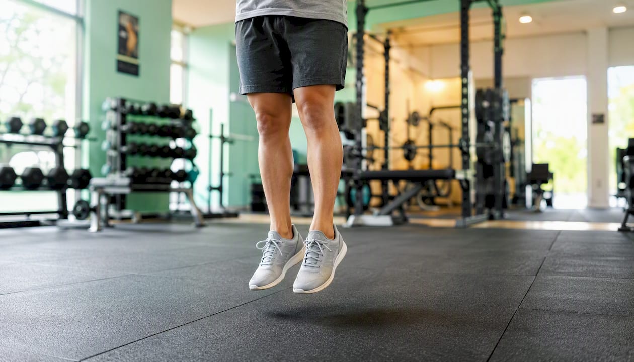
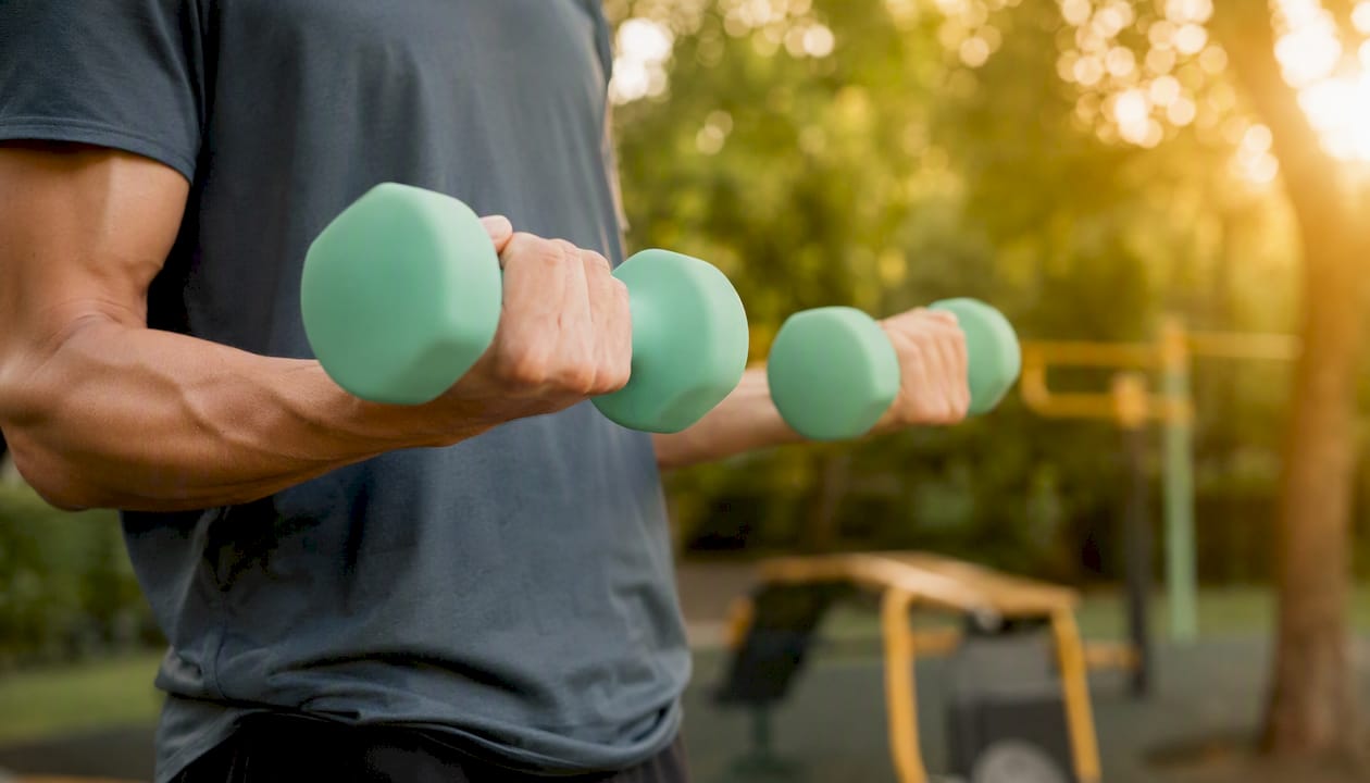
Progressive resistance training is the other non-negotiable. Lifting weights, using resistance bands, or working on machines applies compressive and tensile forces directly to bone. The key word is progressive: the load must increase over time, or the skeleton adapts and stops responding. Two or more strength sessions per week is the NIAMS-recommended minimum for preventing bone loss, and most evidence-backed programs run 3–4 sets of 8–12 reps per major muscle group.
Balance and functional training does not build bone directly, but it prevents the fractures that make bone loss catastrophic. Tai chi, single-leg stands, and heel-to-toe walking reduce fall risk, which matters as much as T-score when you are thinking about long-term skeletal health.
Dose and frequency
NIAMS guidelines recommend adults aim for a moderate amount of aerobic activity each week combined with muscle-strengthening activities at least twice weekly. For bone-specific goals, most exercise physiologists layer in 2–3 impact sessions on top of that base.
Statistic: A 2026 meta-analysis of 22 RCTs covering 1,051 participants found exercise significantly increased lumbar spine BMD (SMD = 0.46) and femoral neck BMD (SMD = 0.42) in young adult women. When exercise modalities were combined, femoral neck BMD improved by SMD = 0.49 and total body BMD by SMD = 0.52 in adult women.
Progression and safety
The 10% rule applies here: increase load, duration, or impact by no more than 10% per week to avoid stress fractures and joint overload. For anyone with low bone mineral density (BMD) or a prior fragility fracture, working with a physical therapist before starting high-intensity resistance training is the safer path. NCBI Bookshelf guidance specifically recommends gradual progression for previously inactive adults.
Red flags — stop and consult a clinician if you experience:
- Sharp, localized bone pain during or after exercise (possible stress fracture)
- Sudden back pain after a low-impact movement (possible vertebral compression fracture)
- Joint swelling that does not resolve within 48 hours
- Dizziness or chest pain during any session
For older adults or those with diagnosed osteopenia, NHS guidance suggests brisk walking, keep-fit classes, and low-impact aerobics as entry points before progressing to higher-impact work. The goal is to find the highest tolerable load, not the lowest comfortable one.
What to eat to support bone density
Bone remodeling is a continuous biological process, and it runs on raw materials. Calcium is the most discussed, but it is not the only one that matters.

Calcium and vitamin D targets
Mayo Clinic and federal guidelines align on these daily calcium targets:
- Adults 19–50: 1,000 mg/day
- Women 51 and older / men 71 and older: 1,200 mg/day
Food sources to hit those numbers: plain yogurt (415 mg/cup), sardines with bones (325 mg/3 oz), fortified orange juice (300 mg/cup), cooked kale (180 mg/cup), and firm tofu made with calcium sulfate (250–750 mg/half cup, depending on brand). Harvard Health Publishing highlights that many people overlook canned fish with bones and fortified plant milks as practical daily sources.
Vitamin D enables calcium absorption in the gut. Without adequate vitamin D, you can hit your calcium target and still absorb very little of it. Most adults need 600–800 IU daily, rising to 800–1,000 IU for those over 70. Fatty fish (salmon, mackerel), egg yolks, and fortified dairy supply some, but few people reach their target through food alone, especially in northern latitudes or with limited sun exposure. Testing serum 25(OH)D is the only way to know where you stand; a level below 20 ng/mL is deficient, and below 30 ng/mL is insufficient by most clinical standards.
Protein, magnesium, vitamin K, and potassium
Higher dietary protein supports BMD by preserving muscle mass, which reduces skeletal stress and fall risk. Aim for 1.0–1.2 g of protein per kilogram of body weight daily. Eggs, legumes, poultry, and Greek yogurt are practical sources that also deliver other bone-relevant micronutrients.
- Magnesium (found in pumpkin seeds, almonds, spinach, and black beans) is a cofactor in bone mineralization; low magnesium is associated with lower BMD in observational data.
- Vitamin K (leafy greens, fermented foods) activates osteocalcin, a protein that anchors calcium into bone matrix.
- Potassium (bananas, sweet potatoes, avocado) helps neutralize dietary acid load, which otherwise draws calcium from bone as a buffer.
- Omega-3 fatty acids (fatty fish, walnuts, flaxseed) have anti-inflammatory effects that may reduce bone resorption, though the evidence is less definitive than for calcium and vitamin D.
A note on high-oxalate foods
Spinach, beet greens, and Swiss chard are nutritious, but their high oxalic acid content binds calcium in the gut and reduces absorption. Eating them alongside other calcium sources is fine; relying on them as your primary calcium source is not. Kale and bok choy have far lower oxalate levels and deliver calcium that absorbs well. The Cleveland Clinic recommends prioritizing low-oxalate leafy greens when building a bone-focused eating pattern.
Pro Tip: Spread calcium intake across two or three meals rather than taking it all at once. The gut absorbs calcium most efficiently in doses of 500 mg or less, so splitting a 1,000 mg daily target into two servings meaningfully improves what actually reaches your bones. For a detailed serving guide, see the foods for bone density resource.
How bone density is measured and who should get tested
What a DXA scan actually measures
Dual-energy X-ray absorptiometry (DXA) is the clinical standard for measuring bone mineral density. It sends two low-dose X-ray beams through bone and calculates how much mineral is present per unit area, reported in g/cm². The result is expressed as a T-score: how many standard deviations your BMD falls above or below the average peak bone mass of a healthy young adult.
Memorial Sloan Kettering Cancer Center notes that BMD reflects both density and structural strength, and that the T-score is the primary tool clinicians use to estimate fracture risk.
| T-score range | Classification | Clinical implication |
|---|---|---|
| Above −1.0 | Normal | Continue preventive lifestyle measures |
| −1.0 to −2.5 | Osteopenia | Lifestyle intervention; monitor every 1–2 years |
| −2.5 or below | Osteoporosis | Clinician review; medication may be indicated |
| −2.5 + fragility fracture | Severe osteoporosis | Specialist referral typically warranted |
Who should get tested and when
Mayo Clinic recommends DXA testing for women soon after menopause and for anyone with significant risk factors. MSKCC advises scanning women within two years of menopause or earlier when specific risks are present.
Get a DXA scan if any of these apply:
- Women aged 65 or older, or men aged 70 or older
- Postmenopausal women under 65 with one or more risk factors (family history, low body weight, smoking, prior fracture)
- Adults who have used oral corticosteroids for three months or longer
- Anyone with a condition associated with secondary osteoporosis (rheumatoid arthritis, celiac disease, hyperparathyroidism)
- Adults who have had a fracture from a low-impact event (a fall from standing height or less)
After a low result
A T-score in the osteopenia or osteoporosis range is not a reason to panic. It is a reason to act. Lifestyle changes — the exercise and nutrition strategies in this guide — are the first-line response for most people with osteopenia. For osteoporosis, a clinician may recommend pharmacotherapy alongside those changes. The two approaches work better together than either does alone.
Lifestyle risk factors and medications that accelerate bone loss
The avoidable risks
Several lifestyle factors actively pull calcium out of bone or suppress the cells that build it.
- Smoking reduces bone-forming cell activity and lowers estrogen levels in women, accelerating loss. Quitting at any age slows the damage.
- Excess alcohol (more than two drinks per day consistently) interferes with calcium absorption and impairs osteoblast function. Moderate intake (one drink per day or fewer) appears neutral.
- Low body weight or a BMI below 18.5 means less mechanical load on the skeleton and often lower estrogen in women, both of which reduce bone stimulus.
- Prolonged inactivity is one of the fastest ways to lose bone. Bed rest studies show measurable BMD loss within weeks.
- High sodium intake increases urinary calcium excretion. Cutting processed food intake is a practical lever here.
- Excessive caffeine (more than four cups of coffee per day) modestly increases calcium excretion, though the effect is small if calcium intake is adequate.
Medications that reduce bone density
Long-term use of certain drugs is a major, underappreciated cause of secondary osteoporosis. The most common offenders:
- Corticosteroids (prednisone, dexamethasone): even three months of daily use can cause significant bone loss; anyone on long-term steroids should discuss bone-protective strategies with their prescriber.
- Proton pump inhibitors (omeprazole, pantoprazole): reduce stomach acid needed for calcium absorption from carbonate supplements.
- Certain anticonvulsants (phenytoin, carbamazepine): accelerate vitamin D breakdown.
- Aromatase inhibitors used in breast cancer treatment: dramatically lower estrogen, causing rapid bone loss.
- Depot medroxyprogesterone acetate (Depo-Provera): associated with BMD reduction during use, though some recovery occurs after stopping.
What to ask your clinician:
- “Does any medication I take affect bone density?”
- “Should I have a baseline DXA given my medication history?”
- “Are there bone-protective alternatives or adjuncts for my condition?”
Bring a complete medication list, your family history of fractures, and any prior DXA results to that appointment.
When supplements help and when prescription therapy is the answer
Supplements with real evidence
Calcium and vitamin D are the two supplements with the strongest evidence base for bone health. When food intake falls short, supplementing makes sense. Calcium carbonate is cheapest but requires stomach acid for absorption, so take it with food. Calcium citrate absorbs without food and suits people on PPIs or with low stomach acid.
Vitamin D3 (cholecalciferol) is more effective than D2 at raising serum 25(OH)D. Doses of 1,000–2,000 IU daily are commonly used to correct insufficiency, but the right dose depends on your baseline blood level. Testing before supplementing is the smarter approach.
Nutraceuticals worth knowing about
- Vitamin K2 (MK-7 form): activates matrix Gla protein and osteocalcin, which direct calcium into bone rather than arteries. Small trials show modest BMD benefits; it is generally safe and pairs well with vitamin D3.
- Magnesium: deficiency is common and associated with impaired vitamin D metabolism. 300–400 mg/day from food or supplements is a reasonable target.
- Creatine: emerging evidence suggests creatine supplementation, combined with resistance training, may support bone mineral density by augmenting muscle mass and training capacity. For a closer look at the evidence, see creatine for bone health.
- Collagen peptides: some trials show modest improvements in bone turnover markers, but the evidence is not yet strong enough to recommend broadly.
Statistic: A 2025 systematic review and meta-analysis found that short-term exercise programs under four months can raise bone formation markers — specifically osteocalcin (OC) and procollagen type 1 N-terminal propeptide (P1NP) — before DXA-detectable BMD changes appear. This means biochemical markers can serve as early signals that your program is working, even when the DXA number has not shifted yet.
Prescription pharmacotherapy
Bisphosphonates (alendronate, risedronate, zoledronic acid) are the most prescribed class for osteoporosis. They slow bone resorption and have a strong fracture-reduction evidence base. Other options include denosumab, romosozumab, and teriparatide, each with different mechanisms, indications, and monitoring requirements.
Prescription therapy is not a replacement for exercise and nutrition. It works best when layered on top of lifestyle changes. Anyone considering pharmacotherapy should have a clinician review their fracture risk using a validated tool such as FRAX before starting.
Safety callouts:
- Bisphosphonates can cause esophageal irritation; take them with a full glass of water and stay upright for 30 minutes.
- Rare but serious risks (osteonecrosis of the jaw, atypical femoral fractures) are associated with very long-term use; most guidelines recommend a drug holiday after 3–5 years for lower-risk patients.
- Never start or stop prescription bone medications without clinician oversight.
Can you actually regain bone density?
The honest answer: yes, but the magnitude depends on your starting point, age, and how consistently you apply the right interventions.
Meta-analysis data give the clearest picture. The 2026 systematic review of 22 RCTs found combined exercise modalities improved total body BMD by SMD = 0.52 in adult women — a statistically significant and clinically meaningful effect. Lumbar spine and femoral neck gains were also significant. These are not trivial numbers; they represent real mineral added to real bone.
Younger adults and those with the lowest baseline BMD tend to see the largest relative gains. Postmenopausal women and older men can still improve, but the ceiling is lower and the timeline is longer. Maintaining what you have becomes as important as rebuilding what was lost.
Statistic: A published case report documents a postmenopausal woman who reversed BMD loss over two years through an integrated program: dietary changes, progressive resistance training, a weighted vest during daily activity, and increased aerobic exercise. Her DXA results improved at both the lumbar spine and femoral neck — without pharmacotherapy.
On timeline: bone formation markers (OC, P1NP) can shift within four months of starting a program. DXA-detectable BMD changes typically take 12–24 months to show up clearly. That gap is not a sign the program is failing. It is how bone biology works. Biochemical markers are the early signal; DXA is the long-term confirmation.
A practical weekly plan to start next week
This schedule combines the exercise categories and nutrition habits that the evidence supports. It is designed for a typical adult with no acute injury or severe osteoporosis. Modify as needed with a physical therapist if you have a T-score below −2.5.
Weekly schedule:
- Monday — Strength training (40–50 min): Squat, deadlift, bent-over row, overhead press, and calf raise. Three to four sets of 8–12 reps each. Rest 60–90 seconds between sets.
- Tuesday — Impact cardio (30 min): Brisk walking with a weighted vest, jogging, stair climbing, or dancing. Aim for moderate-to-vigorous intensity.
- Wednesday — Active recovery or balance work (20–30 min): Tai chi, yoga, or single-leg balance drills. Heel-to-toe walking for 10 minutes counts.
- Thursday — Strength training (40–50 min): Repeat Monday’s pattern or rotate to hip hinge, lunge, lat pulldown, chest press, and plank variations.
- Friday — Impact cardio or sport (30–45 min): Tennis, pickleball, jump rope, or a group fitness class with impact elements.
- Saturday — Longer aerobic session (45–60 min): Hiking, cycling (lower osteogenic value but good for cardiovascular base), or a longer walk with elevation.
- Sunday — Rest or gentle movement: Stretching, a short walk, or nothing. Recovery is part of the stimulus.
Progression for the first three months:
- Weeks 1–2: Establish form and baseline load. Do not push to failure.
- Weeks 3–4: Add 5–10% load to your two primary lifts.
- Month 2: Add a fourth set to your main compound movements.
- Month 3: Introduce one higher-impact session per week (jump squats, box step-ups, or short sprints) if tolerated.
For time-constrained readers, long-term health strategies for high-performing executives offers practical frameworks for fitting structured movement into a packed schedule.
Safety checklist before starting:
- Cleared by a clinician if you have a T-score below −2.5, a prior fragility fracture, or uncontrolled cardiovascular disease
- Proper footwear with grip for impact sessions
- Warm-up of 5–10 minutes before every strength session
- Hydration: at least 16 oz of water before and during exercise
- Stop and seek care for any sharp bone pain, sudden back pain, or chest symptoms
Osteogenic loading and clinic-based therapies: what the evidence says
What osteogenic loading is
Osteogenic loading is a specific protocol that uses specialized equipment to apply brief, high-magnitude compressive forces to the skeleton — forces that can reach multiples of body weight in a controlled, safe setting. This is categorically different from conventional strength training. A typical barbell squat might load the spine at 1–2 times body weight. Osteogenic loading devices are engineered to deliver 4–12 times body weight in a short, isometric-style contraction, targeting the precise mechanical threshold that triggers bone remodeling.
The principle is grounded in Wolff’s Law: bone adapts its structure in response to the loads placed on it. The higher the load (within safe limits), the stronger the remodeling signal. Not all weight-bearing activities are equally osteogenic; combining high-impact loading or applying external load through weighted vests during lower-impact activity markedly increases skeletal stress and the stimulus for new bone formation, as documented in case report evidence.
Who may benefit from clinic-based sessions
Osteogenic loading sessions at a clinic like Osteostrong are particularly relevant for:
- Adults with osteopenia or osteoporosis who want a supervised, measurable bone stimulus beyond what home exercise delivers
- People who cannot safely perform high-impact activities (running, jumping) due to joint pain, balance issues, or prior fracture
- Those who want to complement pharmacotherapy with a targeted mechanical stimulus
- Motivated adults who want to track progress with objective data and professional guidance
Pro Tip: Osteogenic loading is most effective when combined with adequate protein and calcium intake on session days. The mechanical stimulus opens a remodeling window; the nutrients supply the raw material to fill it. If you are curious about what a home-based protocol looks like before committing to clinic sessions, the osteogenic loading at home guide is a practical starting point.
How it fits with other therapies
Osteogenic loading does not replace nutrition, supplements, or prescription medication when those are indicated. It works alongside them. Clinics like Osteostrong also offer adjunct therapies — red light therapy, PEMF mats, vibration plates, and compression therapy — that may support recovery, circulation, and overall musculoskeletal health. The evidence base for these adjuncts varies; red light therapy for bone density and PEMF therapy each have their own developing research profiles worth reviewing before use.
Safety considerations are straightforward: osteogenic loading at a supervised clinic is designed to be safe for people with low BMD, but the protocol should be disclosed to your prescribing clinician, especially if you are on bisphosphonates or have a recent fragility fracture.
When to get medical advice and red flags
Most adults can start a bone-health program without a clinician’s sign-off, but some situations require a medical evaluation first.
Seek care promptly if you experience:
- A fracture from a low-impact event (a fall from standing height, a sneeze, or lifting a light object)
- Sudden, severe mid-back or lower-back pain with no clear mechanical cause (possible vertebral compression fracture)
- Measurable height loss of more than 1.5 inches over time
- Chronic pain in the hip, groin, or thigh that worsens with weight-bearing
Schedule a non-urgent appointment if:
- You have been on oral corticosteroids for more than three months
- You have a first-degree relative who had a hip fracture
- You are postmenopausal and have not had a baseline DXA
- You have a condition associated with secondary bone loss (celiac disease, inflammatory bowel disease, hyperparathyroidism, or hyperthyroidism)
What to bring to your appointment:
- A complete medication list, including supplements and over-the-counter drugs
- Family history of osteoporosis or fragility fractures
- Any prior DXA results, even if from years ago
- A brief log of your current exercise habits and dietary calcium estimate
When a referral to an endocrinologist or a dedicated bone health clinic is reasonable: if your T-score is below −2.5, if you have had two or more fragility fractures, or if your bone loss is progressing despite treatment, a specialist can assess secondary causes and consider therapies beyond first-line bisphosphonates.
The practical path to stronger bones
The conventional framing around bone health tends to be reactive: you get a bad DXA result, and then you start paying attention. What strikes me most about the evidence in this guide is how clearly it argues for the opposite approach.
The 2026 meta-analysis showing significant BMD gains from combined exercise modalities is not describing a pharmaceutical trial. It is describing people who moved with intention and loaded their skeletons progressively. The case report of a postmenopausal woman reversing BMD loss through diet and a weighted vest is not an outlier. It is a proof of concept for what a committed, multi-modal program can do without a single prescription.
What people underestimate is the specificity required. Walking is not enough. Calcium without vitamin D is not enough. Strength training without progressive overload is not enough. Each piece has to be calibrated. The gap between “I exercise and eat reasonably well” and “I am actually stimulating bone remodeling” is wider than most people realize, and that gap is exactly where clinic-based guidance earns its value.
Osteostrong offers a structured path to measurable bone improvement
Diet and exercise are the foundation, and this guide gives you the tools to build on them. For adults who want a supervised, measurable step beyond a home program, Osteostrong offers weekly osteogenic loading sessions that deliver the high-magnitude skeletal stimulus most home workouts cannot replicate. Each session takes about 15 minutes and is calibrated to your current strength and bone health status.
Osteostrong also offers red light therapy, PEMF mats, vibration plates, and hydromassage lounges as adjunct recovery and wellness tools, all under one roof. The model is simple: one session per week, tracked over time, with objective progress data. Adults with osteopenia, osteoporosis, or a strong interest in proactive skeletal health get the most from this format. Find a center near you or visit the Rancho Cordova location to book your first session.
Sources
- 2026 systematic review and meta-analysis (exercise effects on BMD)
- Exercise for Your Bone Health | NIAMS
- Surprising foods that boost bone health
This article provides general health information and is not a substitute for professional medical advice. Consult a qualified clinician to confirm whether these strategies are appropriate for your individual health status and risk profile.