

For bone mineral density, lumbar spine precision errors can be well under 1% and hip precision errors commonly run 1–2% at centers that follow best practices. For body fat, the picture is slightly messier: DXA correlates extremely well with CT (r ≈ 0.99) but can systematically underestimate whole-body fat mass by as much as ~5 kg in some populations.
The single most useful thing to understand before your first scan: for tracking change over time, precision matters far more than absolute accuracy. A DXA machine that is slightly off in absolute terms but highly reproducible will tell you whether you are gaining bone or losing fat far more reliably than a “more accurate” machine with inconsistent positioning protocols.
Key stat: Instrument accuracy error for clinical DXA is often reported as better than 10%, while in-house precision at well-run centers can reach well under 1% for lumbar spine BMD.
Pro Tip: Ask your clinic whether they have performed an in-house precision study and can give you their facility-specific Least Significant Change (LSC) value before you book. If they cannot answer that question, find a different center.
Key Takeaways
| Point | Details |
|---|---|
| Precision beats accuracy for tracking | Reproducibility matters more than absolute accuracy; use the same machine and clinic for every serial scan. |
| LSC is the decision threshold | A change must exceed 2.77 × precision error to be real at 95% confidence; ask your clinic for their facility-specific LSC. |
| Body fat has systematic limits | DXA correlates with CT at r ≈ 0.99 but can underestimate fat mass by up to ~5 kg in some populations. |
| Clinic quality varies widely | Look for ISCD-certified technologists, a published in-house precision study, and daily phantom QC logs. |
| Osteostrong complements monitoring | Weekly osteogenic loading sessions at Osteostrong give you a structured program to act on your DEXA results. |
Table of Contents
- How does DEXA measure body composition and bone density?
- What does the evidence say about DEXA accuracy for body fat?
- How accurate is DEXA for bone mineral density?
- What factors cause DEXA results to vary?
- How do you know if a change between two scans is real?
- How does DEXA compare with other body-composition methods?
- Is DEXA safe, and how often should you get one?
- How to prepare for a DEXA scan and what to ask
- How to pick a clinic with reliable DXA measurements
- What DEXA results actually tell you in practice
- Bone health goes beyond the scan
- Sources
How does DEXA measure body composition and bone density?
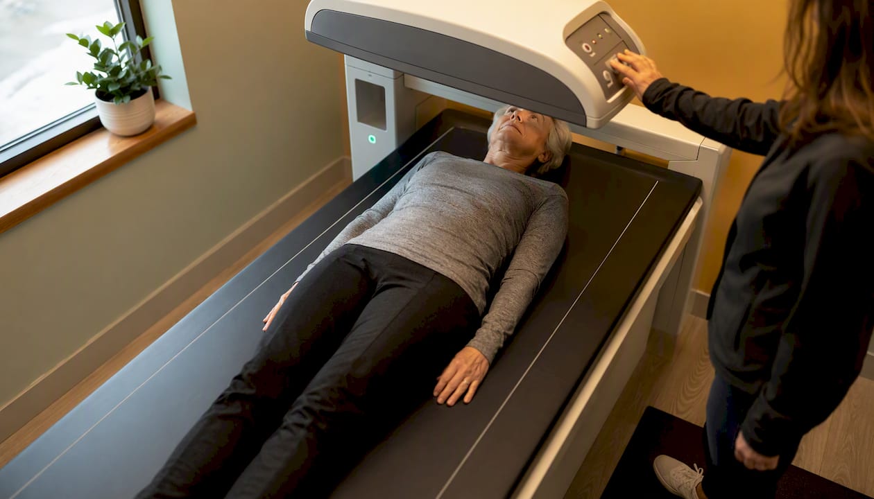
DXA works by passing two X-ray beams at different energy levels through the body and measuring how much each is attenuated by the tissue underneath. Bone, lean soft tissue, and fat absorb the two energies differently, so the scanner’s software can separate them mathematically. The whole-body scan takes roughly 10 minutes and produces a regional breakdown you cannot get from a scale or a blood test.
Typical outputs include:
- Total body fat percentage and fat mass in kilograms
- Lean mass (muscle plus organs, minus bone and fat) by region: arms, legs, trunk, android/gynoid zones
- Bone mineral density (BMD) in grams per square centimeter (g/cm²) at the lumbar spine, total hip, and femoral neck
- T-scores and Z-scores derived from BMD, used to classify osteopenia and osteoporosis against reference populations
Two manufacturer families dominate U.S. clinical practice: Hologic (fan-beam systems, Apex software) and GE Healthcare — Lunar (iDXA and Prodigy systems, enCORE software). Their algorithms differ enough that a BMD value from a Hologic machine is not directly comparable to one from a GE Lunar machine without cross-calibration. Updated practice guidelines are explicit: never compare scans from different machines unless they have been formally cross-calibrated, because even machines of the same model can differ enough that a perceived change is actually measurement noise.
What does the evidence say about DEXA accuracy for body fat?
DXA’s body fat measurements hold up well in controlled research settings but carry real caveats in clinical practice.
Against whole-body CT as a reference, DXA shows high correlation (r ≈ 0.99), which sounds reassuring. The problem is that high correlation does not mean the two methods agree on the absolute number. Systematic differences show up consistently:
- DXA can underestimate whole-body fat mass by up to ~5 kg in some study samples compared with CT, particularly in individuals with higher trunk fat.
- Agreement for total body mass versus a calibrated scale is very close in controlled tests (often within ~1%), confirming the hardware is stable, but the fat/lean partition is where errors accumulate.
- In very obese individuals, scan geometry limits (table width, software assumptions about tissue thickness) can introduce additional error. Older machines had hard weight and width limits; modern systems have improved but extreme cases still require caution.
- In very lean individuals, the two-compartment soft-tissue model DXA uses can overestimate fat percentage because the algorithm has less tissue to work with.
- Older adults with altered hydration and tissue composition represent another group where absolute fat estimates diverge more from reference methods.
Key stat: DXA correlates with CT at r ≈ 0.99 for body composition, yet systematic underestimation of fat mass by up to ~5 kg has been documented in some samples, a gap large enough to matter clinically.
The deeper issue, noted in peer-reviewed analysis, is that absolute validation against chemical analysis (the true gold standard) is limited by the absence of field phantoms that replicate human tissue composition. Interpret absolute fat mass numbers with that caveat in mind. What DXA does exceptionally well is track relative change in the same individual on the same machine over time.
How accurate is DEXA for bone mineral density?
For BMD, DXA operates under a well-defined clinical framework built around the concept of precision, rather than absolute accuracy. The International Society for Clinical Densitometry (ISCD) sets the standard here, and the math is straightforward once you know it.
How precision error and LSC work:
- A facility measures precision error by performing duplicate or triplicate scans on a representative patient sample (ISCD recommends 30 duplicate or 15 triplicate scans per technologist).
- Precision error is expressed as the root-mean-square standard deviation (RMS-SD) in g/cm².
- The Least Significant Change (LSC) is calculated as 2.77 × precision error, representing the threshold a measured change must exceed to be considered real at 95% confidence.
- Serial BMD comparisons use absolute g/cm² values, not T-scores or percent changes alone, because T-scores shift with reference database updates and obscure true longitudinal change.
Key stat: At a center with a low lumbar spine precision error, the LSC is a small value; changes smaller than this cannot be distinguished from measurement noise at 95% confidence.
Typical precision ranges at well-run U.S. ISCD guidance explicitly warn against using manufacturer-supplied precision values in place of a facility’s own in-house study, because real-world operator skill and patient population differ from manufacturer test conditions.
What factors cause DEXA results to vary?
Precision and accuracy both degrade when any of several controllable factors are ignored. The sources of variability fall into three categories.
Patient factors:
- Hydration status: Lean mass and fat estimates shift with hydration because DXA’s algorithm assigns tissue to compartments partly based on X-ray attenuation, which water affects. Scan at the same hydration state each time.
- Recent food intake: A full stomach adds mass and can alter trunk-region soft-tissue readings.
- Metal objects: Jewelry, underwire bras, belt buckles, and implants (joint replacements, spinal hardware) scatter X-rays and corrupt regional data. Implants must be disclosed; some require repositioning or regional exclusion.
- Extreme body size: As noted above, very high body mass or unusual height can push scan geometry toward its limits.
Machine and software factors:
- Hologic and GE Lunar systems use different algorithms; results are not interchangeable without cross-calibration.
- Software version upgrades can shift population reference values, making pre-upgrade and post-upgrade scans incomparable unless the clinic re-runs the analysis.
- System stability with phantom-based QC can be better than 0.5% over long periods for a single well-maintained system, which is why daily phantom scanning matters.
Operator factors:
- Positioning is the largest practical driver of precision error in most clinical settings. A hip rotated even a few degrees changes femoral neck BMD readings measurably.
- Technologist certification and adherence to a written positioning protocol are the main defenses against this.
Pro Tip: For serial body composition or BMD monitoring, always return to the same clinic, the same machine, and request the same technologist when possible. Even switching between two machines of the same model at the same clinic can introduce error that exceeds a real biological change.
How do you know if a change between two scans is real?
This is the question most patients never think to ask, and it is the one that matters most for longitudinal monitoring.
The answer comes from the LSC calculation. Here is how to apply it:
- Get your facility’s precision error for the site being monitored (lumbar spine, total hip, or femoral neck). Ask for it in g/cm² (for BMD) or percentage (for body fat).
- Multiply by 2.77 to get the LSC at 95% confidence.
- Compare the difference between your two scans to the LSC. Only if the measured change exceeds the LSC can you conclude the change is real rather than noise.
Two worked examples:
- If lumbar spine precision error = 1% and your baseline BMD = 1.000 g/cm², the LSC ≈ 2.77%. A follow-up scan showing 1.020 g/cm² (a 2% increase) falls below the LSC. You cannot call that a real gain.
- If precision error = 0.5% at the same site, LSC ≈ 1.4%. That same 2% increase now clears the threshold and represents a statistically meaningful change.
Key stat: If a facility’s precision error for body fat is 2%, the LSC is approximately 5.6 percentage points. A drop from 32% to 28% body fat would not clear that bar. A drop from 32% to 25% would.
ISCD is direct: use the facility’s own LSC, not a manufacturer’s published figure. The difference between a well-trained technologist and a less consistent one can shift the LSC enough to change clinical decisions.
How does DEXA compare with other body-composition methods?
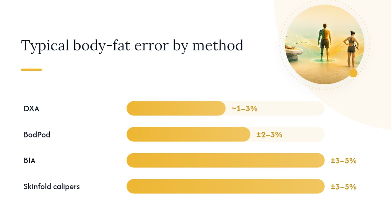
| Method | Typical error range | Main sources of bias | Best use case | Radiation |
|---|---|---|---|---|
| DXA | Body fat: ~1–3% precision; BMD: <1–2% CV | Hydration, positioning, software version, extreme body size | Regional breakdown, serial BMD monitoring | Very low |
| BIA (bioelectrical impedance) | ±3–5% body fat in many studies | Hydration, electrode placement, algorithm assumptions | Screening, home tracking | None |
| Skinfold calipers | ±3–5% body fat (operator-dependent) | Technician skill, site selection, equation choice | Low-cost field assessment | None |
| BodPod (air displacement) | ±2–3% body fat | Clothing, hair, lung volume assumptions | Research, no radiation needed | None |
| CT / MRI | Considered highest accuracy for fat/muscle volume | Cost, availability, radiation (CT) | Research, surgical planning, visceral fat quantification | CT: moderate; MRI: none |
A few practical notes on that comparison:
- BIA is inexpensive and fast but swings widely with hydration. A morning reading after coffee versus an evening reading after a workout can differ by several percentage points on the same device.
- Skinfold calipers depend almost entirely on the technician. In experienced hands they are useful for tracking; in inexperienced hands the error exceeds DXA’s.
- BodPod is a reasonable alternative when radiation is a concern, but it cannot give regional breakdowns or BMD.
- CT and MRI are the reference standards for visceral fat quantification and muscle cross-sectional area, but cost and access limit them to research or specific clinical indications.
Choose DXA when you need regional body composition data, serial BMD monitoring, or a clinically recognized standard for osteoporosis management. Choose CT or MRI when precise visceral fat volume or muscle anatomy is the clinical question.
Is DEXA safe, and how often should you get one?
DXA radiation exposure is exceedingly small, typically less than the natural background radiation you absorb in a single day. For context, RadiologyInfo notes that DXA delivers far less radiation than a standard chest X-ray. The practical safety profile for most adults is excellent.
That said, a short checklist of who should pause or avoid:
- Pregnant individuals: Do not have a DXA scan. Even low-dose radiation carries a precautionary contraindication during pregnancy.
- Recent contrast or nuclear medicine studies: Residual contrast agents can alter soft-tissue attenuation. Most protocols recommend waiting 24–48 hours after contrast studies.
- Metallic implants in the scan field: Not a contraindication, but the affected region may need to be excluded from analysis or the scan repositioned.
- Individuals who cannot lie flat or still: Motion artifact degrades precision significantly.
Key stat: DXA radiation dose is typically very low, generally less than the natural background radiation received in a single day, making it one of the lowest-dose imaging procedures in clinical use.
For BMD monitoring, clinical frequency follows the indication. Patients on osteoporosis therapy are typically rescanned every 1–2 years to assess treatment response, but only after a period long enough that a real change would exceed the facility’s LSC. Rescanning sooner than that produces noise, not signal. Follow your clinician’s guidance on timing.
How to prepare for a DEXA scan and what to ask
Pre-scan checklist:
- Wear loose, metal-free clothing (no underwire, no belt, no zipper at the scan site). Many centers provide a gown.
- Remove all jewelry before arriving, not just in the room.
- Avoid eating a large meal within 2–3 hours of a body composition scan to reduce stomach contents affecting trunk readings.
- Maintain consistent hydration across serial scans. Scan at roughly the same time of day each visit.
- Disclose all implants, joint replacements, spinal hardware, or recent surgeries to the technologist before the scan begins.
- If you have had a recent contrast study (CT with contrast, nuclear medicine scan), tell the scheduling team so they can advise on timing.
Questions to ask the technologist or clinic coordinator:
- “Do you perform an in-house precision assessment, and can you give me your facility’s LSC for lumbar spine and total hip?”
- “Which machine and software version will you use, and is it the same one I used last time?”
- “Are your technologists ISCD-certified or trained to a written positioning protocol?”
- “Will the scan report include the machine ID, software version, and technologist initials?”
- “If my results change between visits, how will you determine whether the change is clinically significant?”
The scan report itself should include: absolute BMD values in g/cm², T-scores and Z-scores with the reference database named, machine model and serial number, software version, and ideally the facility’s LSC or a statement that changes were interpreted against it.
How to pick a clinic with reliable DXA measurements
Not all DXA centers are equal, and the gap between a well-run facility and a poorly run one is large enough to change clinical decisions. Here is what to look for.
Concrete quality signals:
- Technologist certification: ISCD offers the Certified Clinical Densitometrist (CCD) credential; technologists can hold the Certified Densitometry Technologist (CDT) designation. Ask whether staff hold either.
- In-house precision study: The facility should have performed a precision study using 30 duplicate or 15 triplicate scans on a patient population representative of their caseload, per ISCD standards. This is not optional for a quality center.
- Published LSC values: The LSC should be posted or available on request for each skeletal site and each technologist. A center that cannot produce this number is not meeting minimum quality expectations.
- Daily phantom QC: Best-practice guidelines call for daily phantom scanning (or at minimum three times weekly) with logged results. Ask whether the center maintains a phantom QC log and whether you can see a recent plot.
- Named DXA interpreter with credentials: Reports should be signed by a physician or clinician with documented DXA training, not just a generic radiology read.
- Same machine for serial scans: The center should commit to scanning you on the same machine and, where possible, the same software version for all follow-up visits. Cross-calibration between machines is possible but adds complexity; same-machine serial scanning is simpler and more reliable.
Pro Tip: Ask to see the phantom QC chart for the machine you will be scanned on. A well-run center will have a clean, stable trend line. Drift, gaps, or an inability to produce the chart are red flags.
What DEXA results actually tell you in practice
DXA is a genuinely useful clinical tool, but its value depends entirely on how the results are read and what they are read alongside.
A single scan gives you a snapshot: a body fat percentage, a lean mass distribution, a BMD with a T-score. That snapshot is meaningful for diagnosing osteoporosis or establishing a baseline, but it cannot tell you whether you are improving. For that, you need serial scans on the same machine, interpreted against a facility-specific LSC. A change in lumbar spine BMD that looks impressive on paper may fall entirely within measurement noise if the center’s precision error is high.
The other thing a DXA number cannot do is replace clinical context. A T-score of -2.3 means something different in a 55-year-old woman with low calcium intake and a family history of fracture than it does in a 70-year-old man on bisphosphonate therapy for three years. Labs (vitamin D, parathyroid hormone, bone turnover markers), fracture history, fall risk, and medication history all shape what the number means and what to do about it.
The case where this matters most: a patient rescanned after 12 months shows a 0.015 g/cm² increase in lumbar spine BMD. The right response is not to celebrate or to panic, but to continue the current protocol and recheck at the next appropriate interval. That is the kind of interpretation a well-run DXA center should be providing with every report.
Bone health goes beyond the scan
A DEXA scan tells you where your bone density stands. What you do with that information is where the real work begins. Osteostrong is built around osteogenic loading, the specific type of mechanical stimulus shown to trigger bone remodeling, delivered in a single weekly session. Members also have access to red light therapy, PEMF mats, vibration plates, and compression therapy, all in one place.

If your scan results show osteopenia or osteoporosis, or you simply want to build a stronger skeletal baseline before your next BMD test, Osteostrong gives you a structured, science-backed program to act on. Find an Osteostrong center near you and ask about a free introductory session to see how the program fits your bone health goals.
Sources
- Dual-Energy X-Ray Absorptiometry Scanning in Practice, Technical Aspects, and Precision Testing
- Precision Assessment & Calculator FAQs – ISCD
- Updated practice guideline for dual-energy X-ray absorptiometry (DXA)
This article is general information, not a substitute for advice from a qualified doctor. Consult a qualified healthcare professional about your own circumstances before acting on anything here.